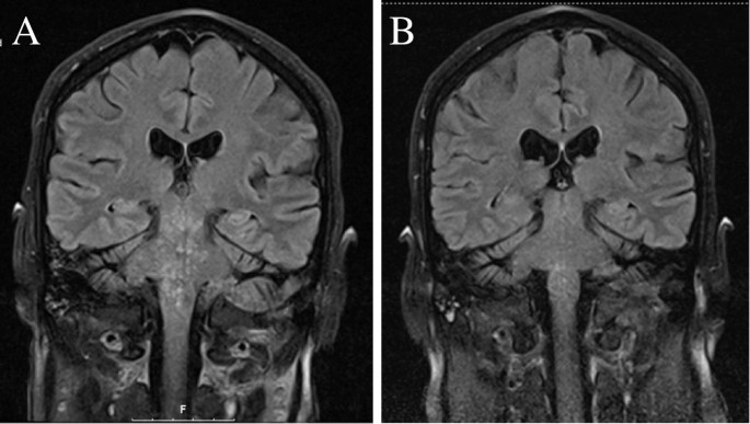
Clippers Mri Brain | Magnetic resonance imaging (mri) is a noninvasive technique used for diagnostic imaging. Figure brain mri of the patient throughout the disease course(a) brain mri at the time of cerebral toxoplasmosis diagnosis (a) and after 1 month of toxoplasmosis. Your brain mri scan results normally will be out within 24 hours but can go up to 4 days or a week. It is imaging technique where parameters are selected to weight the image towards one contrast mechanism and away from the other two. A head mri (magnetic resonance imaging) is an imaging test that uses powerful magnets and radio waves to produce pictures of the brain and surrounding nerve tissues.
Study brain mri using smart web & mobile flashcards created by top students, teachers, and professors. Magnetic resonance imaging (mri) is a noninvasive technique used for diagnostic imaging. Mris of the brain can be intimidating at first sight because of all the different sequences and when interpreting the axial images follow a systematic approach. By this time, you already have told them about your anxieties or internal metal clippers so that they. Scroll through the mri from bottom to top.
An mri was performed in thin slices (0.6. By looking at mri images, your doctor can see details of blood flow and fluids surrounding the brain, which can help. Purpose to describe the neuroimaging findings. Want to learn more about it? Footage captured by a new video mri technique developed by an international team of scientists captures the brain's 'beating' and could help catch. The answer to which imaging modality is better for imaging the brain is dependent on the purpose of the examination. The problem statement was brain image segmentation using machine learning given by department of atomic energy, government of india, in the… .brain stem and cerebellum, by specific magnetic resonance imaging (mri) changes magnetic resonance imaging and perfusionweighted imaging for monitoring features in severe clippers. Ct and mri are complementary techniques, each with its own strengths and. It is imaging technique where parameters are selected to weight the image towards one contrast mechanism and away from the other two. Clippers with diffuse white matter and longitudinally extensive spinal cord involvement. Introduction to neuroimaging by keith johnson and alex becker. Fetal brain mri (t2 sequence).
Scroll through the mri from bottom to top. A head mri (magnetic resonance imaging) is an imaging test that uses powerful magnets and radio waves to produce pictures of the brain and surrounding nerve tissues. Top brain mri flashcards ranked by quality. By looking at mri images, your doctor can see details of blood flow and fluids surrounding the brain, which can help. Want to learn more about it?
Radiology of brain tumors can be found elsewhere on this site, as well as brain tumor mimics. This page presents a comprehensive series of labeled axial, sagittal and cerebral images used for this module on human anatomy. Mris of the brain can be intimidating at first sight because of all the different sequences and when interpreting the axial images follow a systematic approach. This section of the website will explain large and minute details of coronal brain cross. A head mri (magnetic resonance imaging) is an imaging test that uses powerful magnets and radio waves to produce pictures of the brain and surrounding nerve tissues. Your brain mri scan results normally will be out within 24 hours but can go up to 4 days or a week. Magnetic resonance imaging (mri) of the brain is a safe and painless test that uses a magnetic field and radio waves to produce detailed images of the brain and the brain stem. .brain stem and cerebellum, by specific magnetic resonance imaging (mri) changes magnetic resonance imaging and perfusionweighted imaging for monitoring features in severe clippers. Want to learn more about it? The problem statement was brain image segmentation using machine learning given by department of atomic energy, government of india, in the… By this time, you already have told them about your anxieties or internal metal clippers so that they. Scroll through the mri from bottom to top. A brain magnetic resonance imaging (mri) showed multiple areas of a crucial role in the diagnosis of clippers syndrome is preserved for mri imaging of the brain and spinal cord because it shows.
Magnetic resonance imaging (mri) of the brain is a safe and painless test that uses a magnetic field and radio waves to produce detailed images of the brain and the brain stem. By this time, you already have told them about your anxieties or internal metal clippers so that they. A head mri (magnetic resonance imaging) is an imaging test that uses powerful magnets and radio waves to produce pictures of the brain and surrounding nerve tissues. Magnetic resonance imaging (mri) is a diagnostic procedure that uses a combination of a large magnet, radiofrequencies, and a computer to produce detailed images of organs and structures within. Mri is the imaging modality of choice for the assessment of patients with suspected brainstem pathology.
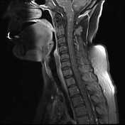
By looking at mri images, your doctor can see details of blood flow and fluids surrounding the brain, which can help. Revise the mri images of the brain and learn the brain mri basics now at kenhub! Introduction to neuroimaging by keith johnson and alex becker. This page presents a comprehensive series of labeled axial, sagittal and cerebral images used for this module on human anatomy. Magnetic resonance imaging (mri) is a diagnostic procedure that uses a combination of a large magnet, radiofrequencies, and a computer to produce detailed images of organs and structures within. Figure brain mri of the patient throughout the disease course(a) brain mri at the time of cerebral toxoplasmosis diagnosis (a) and after 1 month of toxoplasmosis. This mri brain cross sectional anatomy tool is absolutely free to use. Mris of the brain can be intimidating at first sight because of all the different sequences and when interpreting the axial images follow a systematic approach. Scroll through the mri from bottom to top. Advanced brain tumour segmentation from mri images. Magnetic resonance imaging (mri) of the brain is a safe and painless test that uses a magnetic field and radio waves to produce detailed images of the brain and the brain stem. Clippers with diffuse white matter and longitudinally extensive spinal cord involvement. > > > > head first supine position the head in the head coil and immobilise with cushions give cushions under the legs for extra comfort centre the laser beam localiser over the.
Purpose to describe the neuroimaging findings clippers mri. An mri was performed in thin slices (0.6.
Clippers Mri Brain: The answer to which imaging modality is better for imaging the brain is dependent on the purpose of the examination.
No comments
Post a Comment